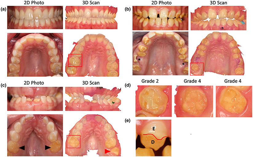
FIGURE 2.
(a) Front view of teeth in Patient A as seen in 2D photo and 3D intraoral scan in the top row. Bottom row shows the occlusal view of the upper (maxillary) teeth in the 2D and 3D views, respectively. The Grade 2 enamel loss was restricted to the molars in upper and lower arch of Patient A. (b) 2D photos and 3D scans of Patient B showing enamel loss affecting all teeth except the incisors. The upper first molars were severely broken (asterisk) and enamel was lost on all surfaces of affected teeth. (c) Patient C had most primary teeth that were unaffected except the second molars (black arrowhead). The erupting first molars (red arrowhead) and incisors had severe enamel loss indicating a Grade 4 enamel defect. (d) Magnified view of the molars marked by red box from Patients A, B, and C showing the severity of enamel loss. (e) Facial surface of canine tooth in Patient B showing partial enamel loss marked by red line. E, enamel, D, dentin

