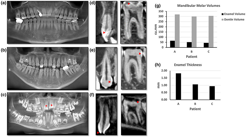
FIGURE 3.
(a–c) Panoramic X-ray reformatted from CBCT. (d–f) Sagittal slice through an anterior (left panel) and one posterior tooth (right panel) from the cone-beam computed tomography scans. (a) Panoramic X-ray of Patient A showing multiple restorations. Enamel is seen as the thin white line outlining the teeth. Several posterior teeth needed restoration due to loss of enamel. (b) Patient B had three restorations visible on the panoramic X-ray and both maxillary first molars have broken crowns (white asterisk) and pulp exposure due to severe enamel loss. (c) Panoramic X-ray of Patient C showing retained primary teeth (white asterisk) and multiple unerupted permanent teeth. The erupting central incisors (red asterisk) seem to have broken incisal edges indicating hypoplastic enamel. (d) Left panel shows 2D slice through anterior tooth in Patient A with normal enamel (red arrowhead). Right panel shows a 2D slice through the molar with thin enamel (red arrowhead). (e) Anterior tooth enamel is irregular and thin on the left panel in Patient B whereas the molar on right panel has no enamel on the occlusal surface and thin enamel on the proximal surface (red arrow heads). (f) Patient C has broken incisal edge with exposed pulp cavity (red arrowhead) of the anterior tooth in left panel. Right panel shows an erupting molar with irregular and patchy enamel seen as white to light gray dots red arrowhead). (g) Bar graph comparing the volumes of enamel and dentin in the three patients. Patients A, B, and C had 65.08, 52.17, and 44.78 cu.mm of enamel, respectively. (h) Bar graph comparing the thickness of enamel in all three patients. The enamel thickness was 1.82, 0.95, and 1.07 mm in Patients A, B, and C, respectively. R: Right, L: Left

