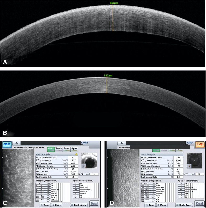
Figure 2.
(A) AS-OCT of the right eye preoperatively shows corneal oedema and increase in pachymetry. (B) AS-OCT of the left eye shows normal findings. (C) Specular microscopy in the right eye shows corneal decompensation with decreased endothelial cells.(D) Specular microscopy in the left eye shows normal morphology and count of endothelial cells. AS-OCT, anterior segment optical coherence tomography.

