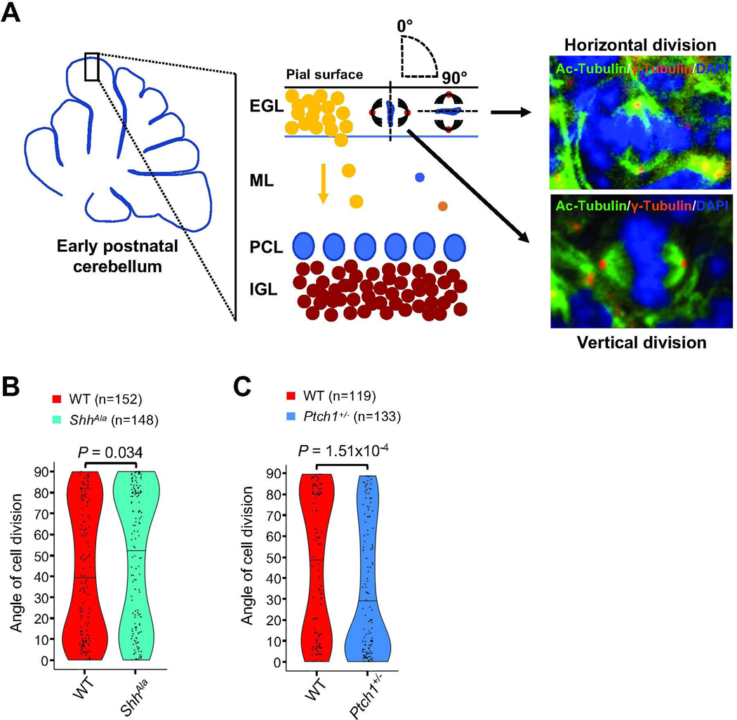
Fig. 1. Shh signaling regulates spindle orientation of mitotic GCPs.
(A) Schematic illustrating the cerebellar cortex and main cell types during early postnatal development. Angles of division for vertically (angle of 0°) and horizontally (angle of 90°) dividing GCPs in the EGL (external granule cell layer) are depicted in the schematic, and representative immunostainings at P3 from sagittal sections in the vermis visualizing both centrioles (γ-Tubulin) and spindles (Ac-Tubulin) of mitotic GCPs in anaphase with the plane of division oriented vertically and horizontally with respect to the pial surface are shown. ML: molecular layer; PCL: Purkinje cell layer; IGL: internal granule cell layer. (B) Violin plots illustrating the distribution of angles of division of mitotic GCPs in the EGL of ShhAla mice at P3 as compared to wild type (WT) mice (n = 5 for each condition, Mann-Whitney test). (C) Violin plots illustrating the distribution of angles of division of mitotic GCPs in Ptch1+/− mice at P3 as compared to WT mice (n = 5 for each condition, Mann-Whitney test).

