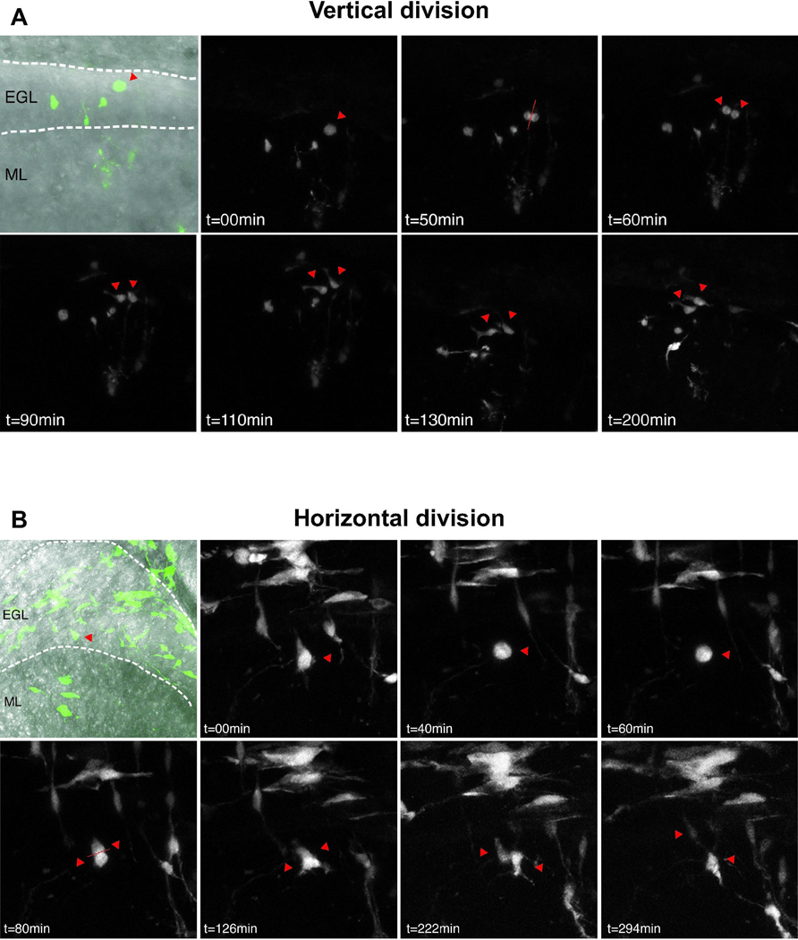
Fig. 2. Live imaging of retroviral GFP labeled GCPs.
Representative images of individual, GFP-labeled GCPs imaged every 10 minutes for up to 24 hours. At P3, 25 divisions were captured out of 101 cells imaged from 4 slices; at P6, 10 divisions were captured out of 110 cells imaged from 5 slices. Single red arrowheads indicate cells before mitosis and double arrowheads indicate position of daughter cells. Dotted lines indicate boundaries of the EGL and IGL. (A) In vertical divisions, both daughter cells remain in the EGL and maintain GCP morphology with short processes. (B) During horizontal divisions, one daughter cell remains in the EGL while the other one migrates into the IGL and exhibits bipolar migratory neuron morphology.

