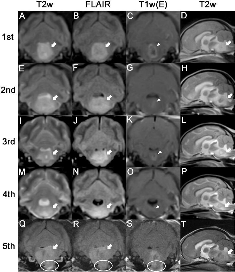
Figure 1.
Serial MRI characteristics of astrocytoma in an 8-year-old neutered male Yorkshire Terrier dog. The first four MRIs were performed using a 0.3-Tesla unit, and the fifth scan was performed using a 1.5-Tesla unit. (A-D) The first MRI scan was acquired before chemotherapy. The tumor lesion (arrows) showed hyperintensity on T2-weighted and fluid-attenuated inversion recovery images. Postcontrast image exhibited ring enhancement (arrowhead). (E-H)Second MRI taken 30 days after chemotherapy. A larger area of peritumoral edema (arrows) compared with that observed on the first MRI scan was observed, and a reduction in the size of the contrast-enhanced lesion (arrowhead) was observed. (I-L) Third MRI scan (taken on day 213). The peritumoral edema (arrows) decreased compared with previous MRI observations, and the contrast enhanced mass (arrowhead) slightly increased in size. (M-P) Fourth MRI scan (taken on day 569). While the peritumoral edema (arrows) increased to a slight extent compared with the observation from the third MRI scan, contrast enhancement (arrowhead) was rarely observed. (Q-T)Fifth MRI scan (taken on day 1155). A much larger tumor (arrows) than those observed on previous MRI scans was noted, and protrusion of the tumor (circles) into the nasopharynx was also observed.

