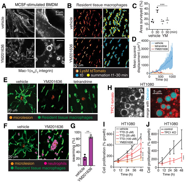
Figure 4. Vacuolar resolution maintains cellular responsiveness and tissue surveillance.
A, BMDM were stimulated for 30 min with M-CSF, with or without YM201636, fixed and immunostained for Mac-1. B-E, Resident tissue macrophages (LysM-tdTomato) with or without YM201636 or tetrandrine were stimulated with M-CSF for 10 min followed by removal of the stimulus for 30 min. Cells were then imaged for 30 min. C, surveillance area measured in the absence (left) or presence (right) of YM201636 (YM) over time, n=3. In D-E, 30 min after M-CSF removal, laser-induced microlesions were generated, marked by the resultant autofluorescence (orange in E). Mean squared displacement of the macrophages is graphed in D; means ± SD, n=3. Representative images in E taken at 15 min post-injury. F-G, Experiments performed as in E. Representative images denoting the presence of neutrophils are shown. In G, the % of lesions with neutrophil swarming was quantified for 6 microlesions per animal, n=3. H, HT1080 cells expressing TPC1-tomato were incubated with 70 kDa dextran for 10 min before imaging. I-J, wildtype and TPC1 KO HT1080 cell growth measured in the absence or presence of YM201636 or tetrandrine (TTD) by cell counting. Means ± SD, n=3. All p values determined by Mann-Whitney U tests.

