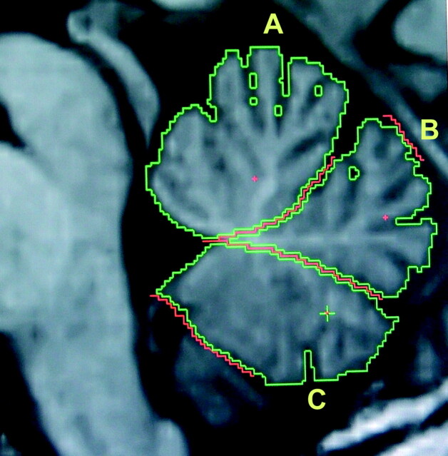
Fig 1.
Sample manual region-of-interest tracings assessing planimetric measurements of the vermal-functional areas by using the MRreg software.14 Data are shown for a control subject with no atrophy. A, The anterior lobule. B, The posterior superior lobule. C, The posterior inferior lobule. A + B + C = midsagittal vermal area.

