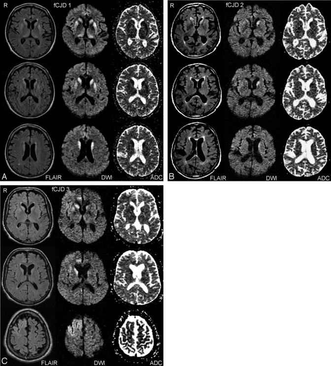
Fig 1.

Representative imaging findings on FLAIR, DWI, and ADC sequences in 3 different subjects with familial CJD. In A (fCJD 1), signal intensity abnormality involves the caudate nucleus, putamen, and thalamus, bilaterally. B (fCJD 2) demonstrates abnormal signal intensity primarily in the left caudate nucleus. In C (fCJD 3), there is involvement of both caudate nuclei, right greater than left, right cingulate gyrus, and right frontal lobe.
