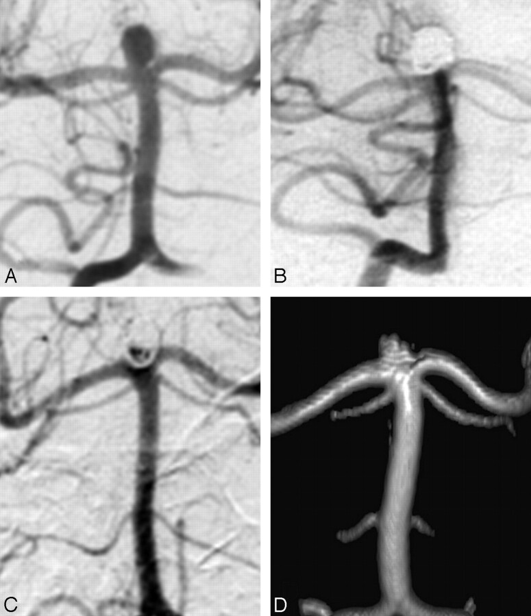
Fig 3.
A 54-year-old woman with a ruptured 5-mm basilar tip aneurysm. A, Precoiling vertebral angiogram shows a basilar tip aneurysm. B, Stable complete occlusion at 6 months. C, Angiogram at 5.8 years shows a 2 × 3 mm disklike recurrence at the base of the aneurysm. D, MRA at 8.0 years is unchanged, compared with the last angiogram.

