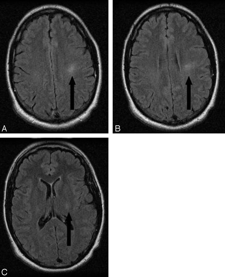
Fig 1.
Axial FLAIR images (TE = 140 ms; TR = 11,002; inversion time = 140 ms) obtained at levels of the centrum semiovale (A), corona radiata (B), and posterior limb of the internal capsule (C) demonstrate increased T2 signal intensity within the left corticospinal tract 5 months after the electrocution injury.

