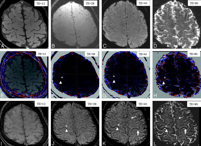
Fig 1.
Comparison of various TEs in the 3 different techniques (magnitude, phase, and phase-weighted imaging) performed in a healthy volunteer (a 37-year-old man): magnitude images with TEs of 12 (A), 28 (B), 40 (C), and 80 ms (D); phase images with TEs of 12 (E), 28 (F), 40 (G), and 80 ms (H); and phase-weighted images with TEs of 12 (I), 28 (J), 40 (K), and 80 ms (L). The gray-to-white-matter contrast on magnitude images (A–D) is poor in all TEs. The gray-to-white-matter contrast on the phase (E–H) and phase-weighted images (I–L) increases by increasing the TE (asterisk versus arrowhead). The heightened contrast between motor (large arrow) and other cortices (small arrow) is most apparent on the phase-weighted image with a TE of 40 ms (K). The definition between motor (large arrow) and other cortices (small arrow) is poor on the phase-weighted image with a TE of 80 ms (L), due to the decrease in signal intensity of the other cortices.

