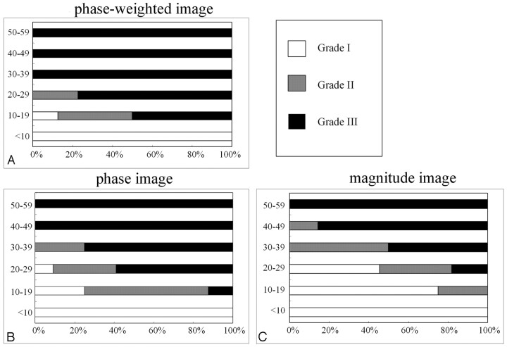
Fig 2.
Distribution of signal intensity in the motor cortex according to the patient's age on phase-weighted (A), phase (B), and magnitude images (C). In all subjects older than 20 years, the motor cortex on phase-weighted images was classified as grade II or III. The frequency of grade III on a phase-weighted image was higher than that on the phase and the magnitude images.

