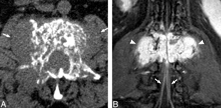
Fig 1.
A, Noncontrast CT demonstrates mixed trabecular and lytic infiltration of the L2 vertebra by a soft tissue mass, expanding the right pedicle and extending into paravertebral soft tissues (arrows). B, Postgadolinium T1 fat-sat coronal demonstrates the avidly enhancing, lobulated mass (arrowheads) with prominent flow voids. Note enhancement of the cauda equina below the level of compression (arrows).

