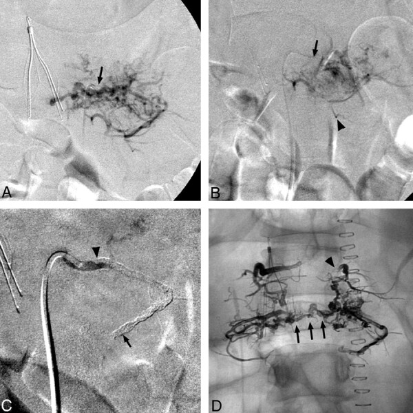
Fig 2.

A, Selective angiography of the left L2 lumbar artery shows prominent tumor vascularity. No spinal anastomoses were identified. Note inferior vena-cava filter and diagnostic catheter in situ (arrow). B, Post-Embosphere-embolization angiography via the Prowler microcatheter (Cordis) (arrowhead) reveals persistent tumor enhancement. C, Note the guide catheter (arrow) after Onyx embolization, where the artery is occluded but the agent is largely restricted to the main artery (extending from arrowhead to arrow). D, Plain anterior/posterior radiograph shows distribution of the 4 Onyx embolizations. Note extension of Onyx from the proximal left L1 lumbar artery (arrowhead) inferior to L2 and then across the midline (arrows).
