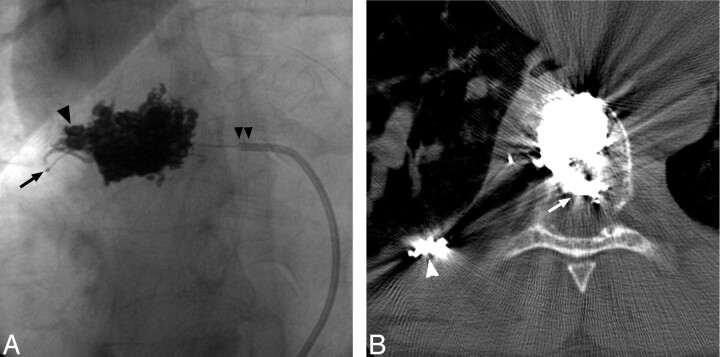
Fig 4.
Postembolization frontal radiograph of the T8 vertebra (A) shows Onyx distributed diffusely through the midlateral and right-lateral vertebral body, extending into the right pedicle (arrowhead). Note tip of Cobra catheter (Merit Medical Systems, South Jordan, Utah) (double arrowheads) and microcatheter positioned in tumor feeder (arrow). Noncontrast CT through T8 shows that the Onyx also extends into the epidural portion of the mass (arrow). Note coils positioned more distally in the right T8 intercostal artery (arrowhead).

