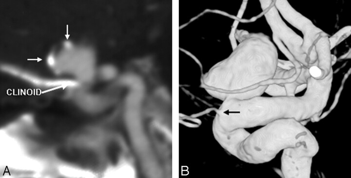
Fig 3.
A 78-year-old woman with SAH from an aneurysm more than 10 mm in size. MSCTA showed a 12-mm aneurysm in the periophthalmic ICA segment on 3D-VR images (data not shown), with peripheral calcifications, best seen on MPR (short white arrows, A). The aneurysm was noted to be separate from the ophthalmic artery origin on 3DRA (black arrow, B).

