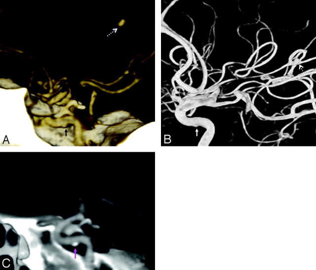
Fig 7.
Atherosclerosis simulating a blister-like lesion. A 52-year-old woman with SAH and focal parenchymal hematoma on CT. CTA showed a 2.9-mm M3 mycotic aneurysm adjacent to the hematoma, also present on 3DRA (dashed arrows, A and B). However, a sessile outpouching was noted on the cavernous ICA undersurface (solid arrows, A and B). Further review of the CTA MPR images revealed this to be an atherosclerotic calcification (pink arrow, C).

