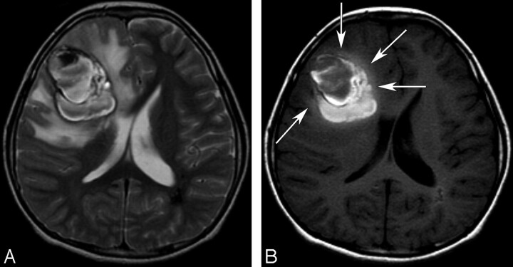
Fig 2.
An 11-year-old boy with a cavernous angioma. A, An axial T2-weighted image (TR/TE/NEX, 3683/104/2 ms) shows a large hemorrhagic mass with heterogeneous signal intensity and peripheral hypointense rims in the right frontal lobe. A perilesional massive edema and mass effect is seen. There is a hypointense lesion in the periventricular white matter of the left parietal lobe and a few small hypointense lesions that were also found in both hemispheres on a T2* gradient-echo image (data not shown), which indicate possible multiple cavernous angiomas. B, An axial T1-weighted image (TR/TE/NEX, 500/9/2 ms) shows a T1 hyperintense perilesional signal intensity sign and mild hyperintensity of a perilesional edema at the deep area abutting the hemorrhagic mass (arrows). Note the centripetal pattern of the T1 hyperintense perilesional signal intensity sign in which T1 hyperintensity within the perilesional edema is observed only in the deep area around the hemorrhagic mass; it is not observed at the periphery of edema.

