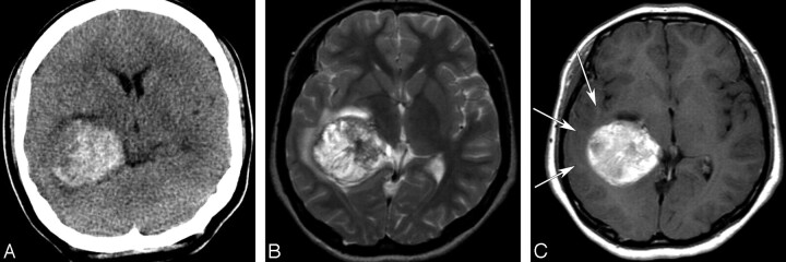
Fig 3.
A 17-year-old girl with a glioblastoma. A, An unenhanced CT image shows a hyperattenuated acute hemorrhagic mass in the right temporal lobe. B, An axial T2-weighted image (TR/TE/NEX, 5000/96/2 ms) shows a hemorrhagic mass with heterogeneous signal intensity and a mild peritumoral edema. C, An axial enhanced T1-weighted image (TR/TE/NEX, 500/12/1 ms) demonstrates mild hypointensity or isointensity of the peritumoral edema (arrows) and mild heterogeneous enhancement of the hemorrhagic mass, which was hyperintense on an unenhanced T1-weighted image (data not shown).

