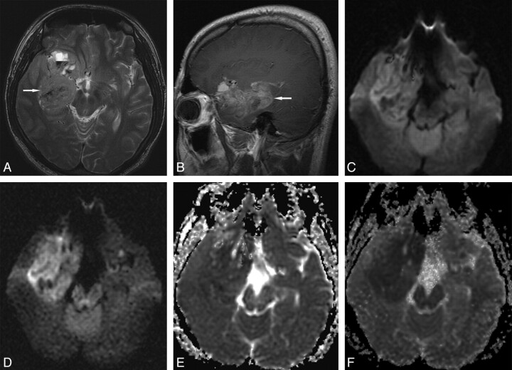
Fig 1.
Grade IV glioblastoma in a 27-year-old man. A, Transverse T2-weighted image shows a slightly hyperintense main mass (arrow) in the right medial temporal lobe. B, Sagittal contrast-enhanced T1-weighted image shows diffuse tumor enhancement (arrow). C, Transverse DWI at b = 1000 s/mm2 shows slight tumor hyperintensity with some hypointense foci. D, Transverse DWI at b = 3000 s/mm2 shows more conspicuous main mass hyperintensity. E, Transverse ADC map at b = 1000 s/mm2 shows subtle or slight tumor hypointensity. F, Transverse ADC map at b = 3000 s/mm2 shows more conspicuous tumor hypointensity.

