Fig 1.
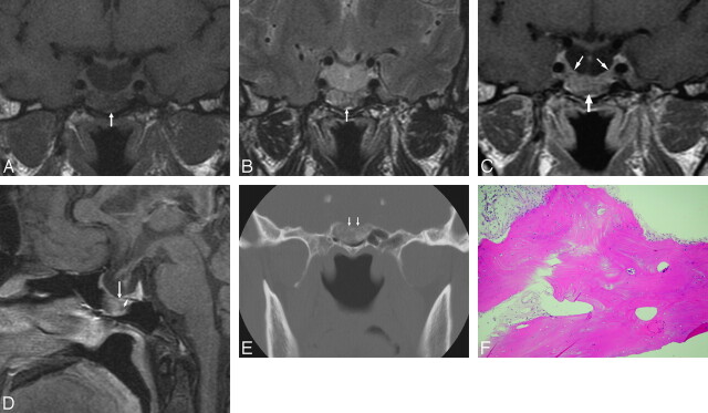
A 50-year-old woman with a sphenoid sinus osteoma in the sella turcica associated with empty sella. A, Coronal T1-weighted (366.7/13.9 [TR/TE]) image of the sella shows a mass lesion (arrow) with low signal intensity, near the pituitary gland, at the sella turcica floor protruding into the sphenoid sinus. Note the associated empty sella. B, Coronal T2-weighted (3000/105 [TR/TE]) image at the same level shows a high-signal-intensity change of the mass (arrow). C, Postgadolinium coronal T1-weighted (750/9.6 [TR/TE]) image shows heterogeneous and moderate enhancement of the mass (arrow), similar to that of the adjacent normal pituitary gland (thin arrows). D, Postgadolinium sagittal T1-weighted (800/11.7 [TR/TE]) image shows a flattened pituitary gland and the sellar floor mass (arrow) protruding into the sphenoid sinus. The dark line of the sellar floor (arrowhead), indicating the intact bony cortex, cannot be detected between the pituitary gland and the tumor, comparing the adjacent sellar floor with intact bony cortex. E, Coronal CT scan of the sella with a bone window setting shows linear calcification within the sellar floor mass. The cortical line of the sella turcica floor (arrows) is involved. F, Photomicrograph of the specimen obtained from incisional biopsy. The tissue is made up of hyperplastic lamellar bone, compatible with osteoma (hematoxylin-eosin, original magnification ×100).

