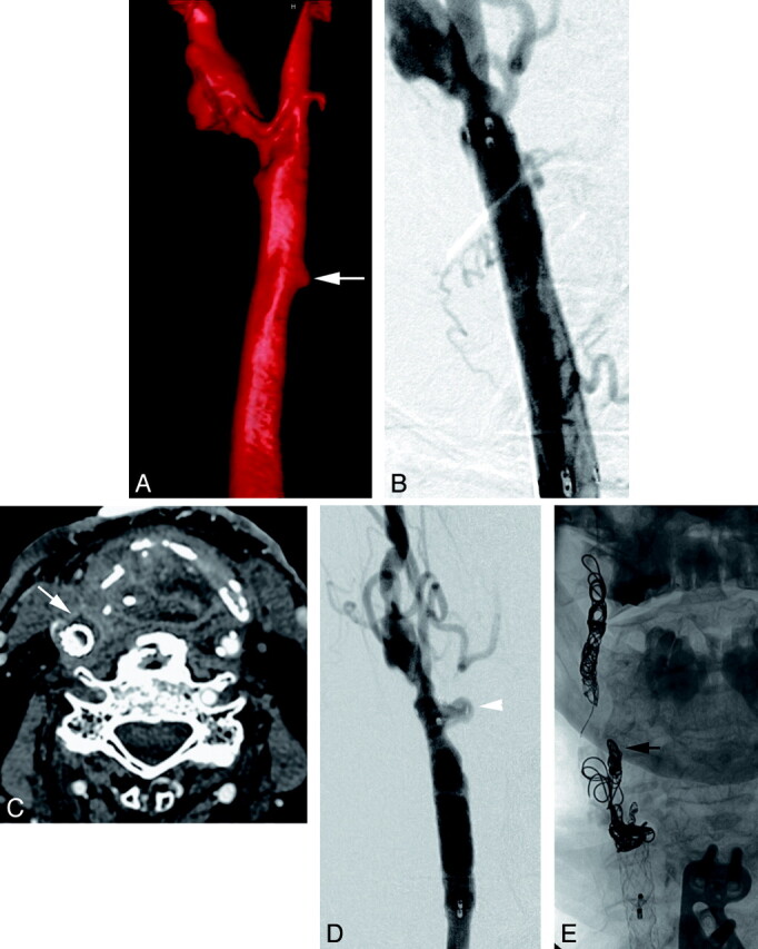
Fig 7.

A 65-year-old patient with a previously treated supraglottic laryngeal carcinoma presenting with profuse hemoptysis. A, A volume-rendered 3D digital subtraction angiogram reveals a small pseudoaneurysm (arrow) arising from the medial aspect of the right common carotid artery. Also note the atherosclerotic changes at the carotid bifurcation. B, This image was obtained following deployment of an 8 × 40 mm stent-graft in the common carotid artery and excluding the pseduoaneurysm. C, The patient presented 11 weeks later with a recurrent hemorrhage from his tracheostomy site. Neck CT shows a rim-enhancing fluid collection (arrow) adjacent to the stent-graft suggestive of infection. D, Common carotid angiogram at this time shows a recurrent carotid blowout (arrowhead) at the distal end of the stent-graft. E, Following a successful temporary balloon occlusion test, the ICA and ECA (black arrow) were occluded with coils, and the common carotid artery was sacrificed proximal to the pseudoaneurysm. The patient has not had another bleed in the following 5 months.
