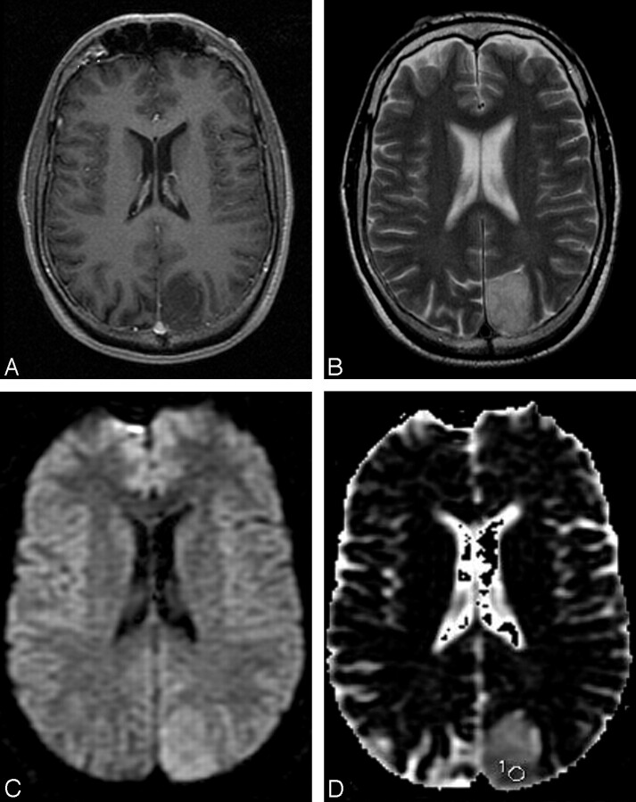
Fig 3.
A 44-year-old man with a grade II diffuse astrocytoma. A, The contrast-enhanced T1-weighted image shows a nonenhancing mass in the left parietal lobe. B, The mass has relatively homogeneous increased signal intensity in the T2-weighted image. C, The DWI shows increased signal intensities compared with the surrounding regions. D, The ADC map shows high a ADC value compared with surrounding brain parenchyma with a subtle low ADC area with a region of interest placed for the lowest ADC measurement. The measured minimum ADC within the mass is 1.185 × 10⁻³ mm2 · sec−1.

