Fig 1.
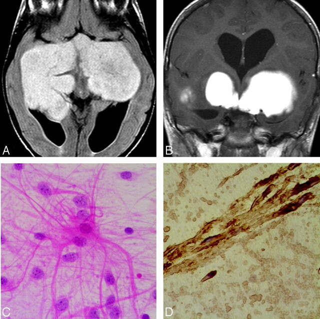
Case 9. A, Axial FLAIR sequence in a 3-year-old girl with progressive blindness shows a large solid lobulated suprasellar and bitemporal PMA, with uniform hyperintensity. B, Coronal contrast-enhanced T1-weighted image shows a homogeneously enhancing suprasellar and bitemporal PMA. C, Photomicrograph shows classic hairlike (“piloid”) astrocytes in a myxoid background (hematoxylin-eosin, original magnification ×300). D, Photomicrograph shows that the tumor is strongly positive for glial fibrillary acidic protein, confirming its astrocytic origin.

