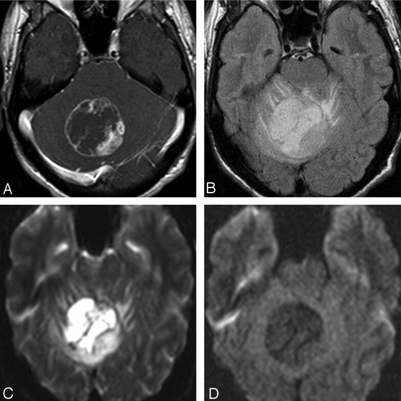
Fig 3.
Case 13. A, Axial contrast-enhanced T1-weighted image in a 17-year-old boy with intractable headaches shows a rim-enhancing PMA of the cerebellar vermis. B, FLAIR image shows heterogeneous hypertintensity with extension into adjacent white matter of the cerebellar folia. C and D, DWI (C) and apparent diffusion coefficient map (D) show no restricted diffusion. This case demonstrates atypical age, location, and enhancement pattern.

