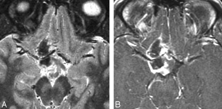
Fig 5.
Case 20. A, Axial T2WI in a 21-year-old man with headache shows a heterogeneous supra- and juxtasellar mass. The hypointense core suggests hemorrhage, which was confirmed at surgery. B, Axial contrast-enhanced T1-weighted image shows a rim-enhancing tumor surrounding a nonenhancing hemorrhagic core. Hemorrhagic exophytic hypothalamic/chiasmatic tumor was partially resected and originally diagnosed as PA in 1997. Re-examination of the specimen in 2007 showed features consistent with PMA.

