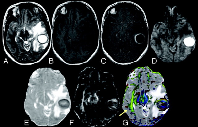
Fig 1.
A 31-year-old female patient presenting with pyogenic brain abscess in the left temporal lobe. A, Axial T2-weighted image shows a well-defined hyperintense lesion with a hypointense wall. B, The lesion appears hypointense on the axial T1-weighted image with an isointense wall. C, On the postcontrast T1-weighted image, the lesion shows ring enhancement. D, DWI shows homogeneous hyperintensity in the cavity that appears hypointense on the MD map (E). The FA (F) and red-green-blue color-modulated FA map fused with the MD map (G) show that high FA in the abscess cavity is similar to what is observed in the contralateral inferior longitudinal fasciculus and midbrain (arrow).

