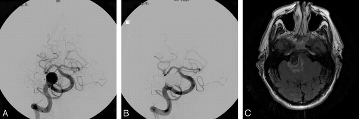
Fig 2.
A, Pre-embolization angiogram showing a large basilar apex region aneurysm. B, Postembolization angiogram showing the coiled aneurysm with minimal residual neck. A large posterior communicating artery on the left produces washout of the left posterior cerebral artery. C, Axial flare MR image showing edema within the brain stem adjacent to the coiled aneurysm.

