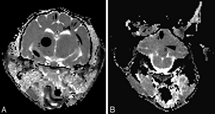
Fig 1.
A and B, ADCav maps of the brain at the level of the thalamus (A) and the cerebellum (B) are shown. The brain regions for which data were acquired include 4 gray matter regions (1 indicates cerebral cortex; 2, thalamus; 3, hippocampus; 4, cerebellar gray matter) and 4 white matter regions (5 indicates corona radiata; 6, centrum semiovale; 7, internal capsule; 8, cerebellar white matter).

