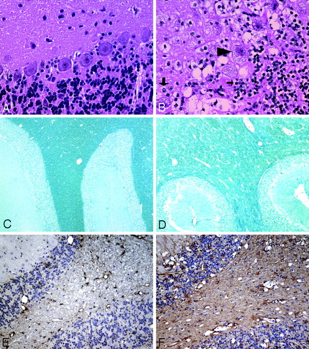
Fig 4.

A and B, H&E-stained sections of the cerebellar cortex (original magnification ×400) from a normal cat (A) and an AMD cat (B), showing cytoplasmic vacuolation and distention of Purkinje cells (arrowhead), granular cells (small arrow), and glia (large arrow). C and D, Luxol fast blue staining of the cerebellar white matter (original magnification ×50) from a normal cat (C) and an AMD cat (D). A decrease in myelination is seen as a decrease in stain intensity and as an increase in space between myelin sheaths. E and F, GFAP immunohistochemistry of the cerebellum (original magnification ×200) from a normal (E) and an AMD cat (F), showing diffuse astrogliosis (brown staining) present throughout the gray and white matter of affected cats.
