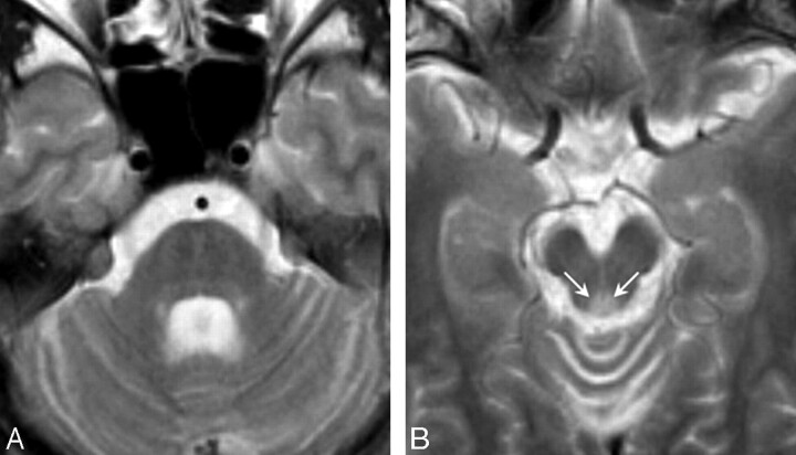
Fig 1.
T2-weighted axial images in patient 1 at 22 years of age (disease duration, 14 years). A, The mid pons and midbrain are slightly atrophic, and the middle and superior cerebellar peduncles show mild atrophy. B, The periaqueductal gray matter signal intensity is abnormally hyperintense (arrows).

