Fig 2.
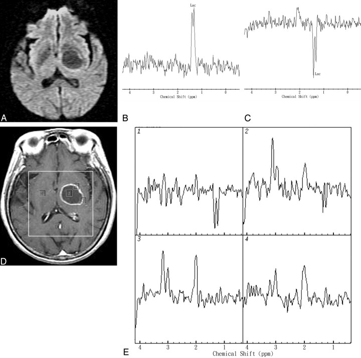
Representative in vivo MR images and spectra from a 67-year-old man with a pathologically proved left thalamus region GBM. A, DWI revealed hypointensity and increased ADC values in the range of 2.51–2.65 × 10⁻³ mm2/s in the cavity of the mass lesion. B and C, In vivo proton single-voxel spectra with a point-resolved spectroscopy sequence (1600/272 ms and 136 ms) from the VOI centered within the cystic mass lesion show a Lac peak at 1.3 ppm that is inverted at a TE of 136 ms. D, Axial contrast-enhanced T1-weighted MR image (500/30) shows a ring-enhanced lesion in left thalamus region and the area of the spectroscopy measurement (VOI) on MR spectroscopic imaging. Voxels corresponding with the center and enhancing rim of the lesion and corresponding contralateral normal-appearing deep gray matter are highlighted in the contrast-enhanced T1-weighted MR image. The spectra were acquired with TE = 136 ms and nominal spatial resolution at 1 mL. E, The spectra from those voxels are shown in detail. The spectra show Lac peak in the center, increased Cho/Cr ratio (maximum, 2.43), and increased Cho/Cho-n ratio (maximum, 2.54) in the rim-enhancing areas of the mass lesion. The contralateral normal-appearing deep brain region does not show any spectral alterations.

