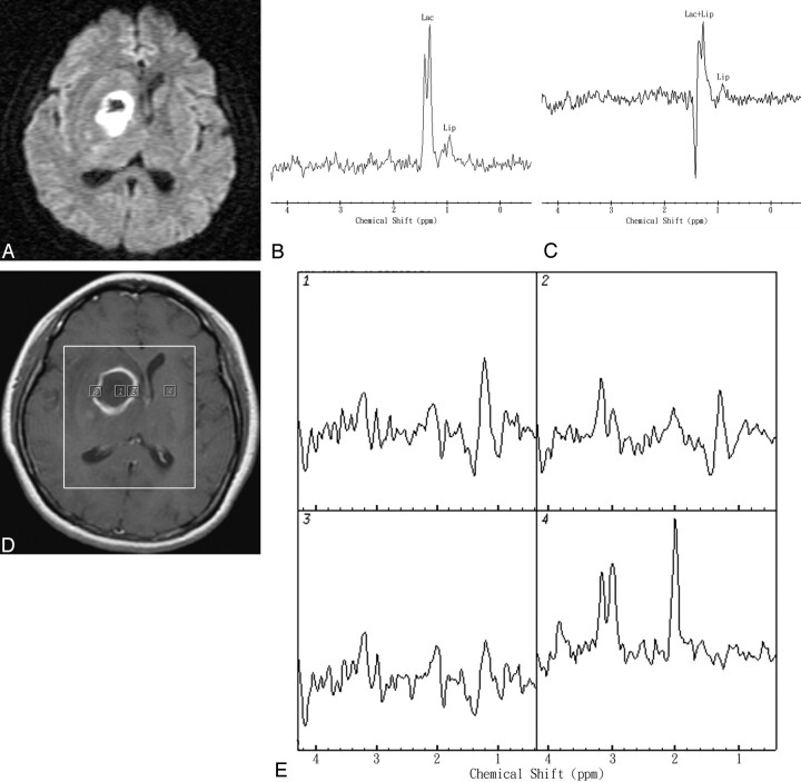
Fig 3.
Representative in vivo MR images and spectra from a 65-year-old woman with stereotactic aspiration proved pyogenic brain abscess in the right deep basal ganglion region secondary to S aureus (facultative aerobe) infection. A, DWI revealed heterogeneous hyperintensity and reduced ADC values in the range of 0.48–0.71 × 10⁻³ mm2/s in most of the cavity. B and C, In vivo proton single-voxel spectra with point-resolved spectroscopy sequence (1600/272 ms and 136 ms) from the VOI centered within the cystic mass lesion show Lac signal intensity at 1.3 ppm, which is inverted at a TE of 136 ms and Lip signal intensity at 0.9–1.3 ppm. D, Axial contrast-enhanced T1-weighted MR image (500/30) shows a rim-enhanced lesion in the right basal ganglion region and the area of the spectroscopy measurement (VOI) on MR spectroscopic imaging. Voxels corresponding with the center and enhancing rim of the lesion and corresponding contralateral normal-appearing deep gray matter are highlighted in the contrast-enhanced T1-weighted MR image. E, The spectra from those voxels are shown in detail. The spectra show Lac and Lip peaks in the center, mild increased Cho/Cr ratio (maximum, 1.82), and decreased Cho/Cho-n ratio (maximum, 0.85) in the rim-enhancing areas of the mass lesion. The contralateral normal-appearing deep brain region does not show any spectral alterations.

