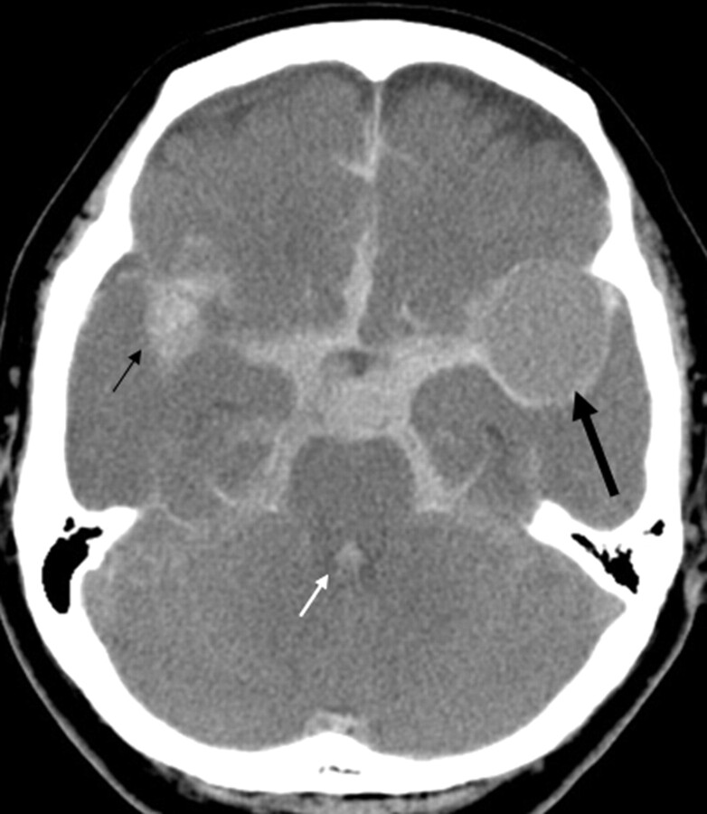
Fig 5.
A 72-year-old woman of the SAH-CPA group. She suddenly fell unconscious and experienced CPA. The first-day CT shows diffuse HDAs in the basal cisterns, Sylvian valleculae/fissures, and cerebral sulci. Note hematoma in the right Sylvian vallecula (small arrow) and in the fourth ventricle (white arrow). The brain shows diffuse subtle low attenuation with obliteration of the cerebral sulci. The CT values of the HDA and the brain are 49 and 29 HU, respectively. In the left Sylvian vallecula, there is a round filling defect in the hematoma that is thought to represent an aneurysm (large arrow).

