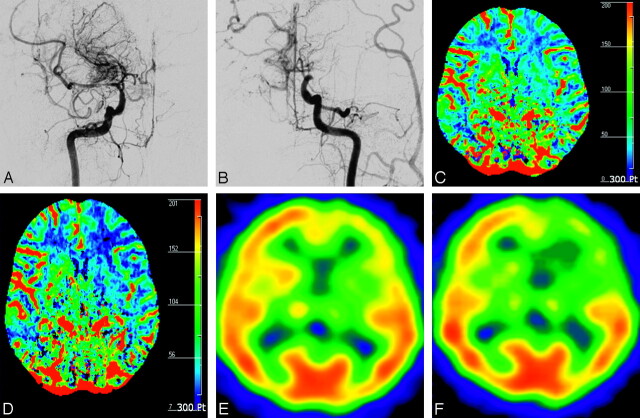
Fig 2.
A 48-year-old woman with transient ischemic attack (case 16). A, Anteroposterior view of right internal carotid arteriogram shows severe stenosis in the proximal M1 portion of MCA with developed BMVs (modified Suzuki stage II). B, Anteroposterior view of left internal carotid arteriogram shows occlusion of distal ICA without antegrade flow (modified Suzuki stage IV). CBF maps before (C) and after (D) ACZ administration show decreased CVR in the left anterior EBZ (white arrow) and MCA territory (black arrow) ipsilateral to the hemisphere with higher modified Suzuki stage compared with contralateral hemisphere with lower modified Suzuki stage. Corresponding SPECT before (E) and after (F) ACZ administration show decreased CVR in the same areas.

