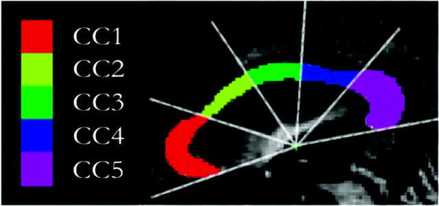
Fig 2.
Segmentation and subdivision of the CC area into 5 distinct subregions obtained from the normalized midsagittal T1 (MPRAGE) scans. A radial partitioning scheme is used for regional analyses of the CC. CC1 indicates rostrum and genu; CC2, rostral body; CC3, midbody; CC4, isthmus; CC5, splenium.

