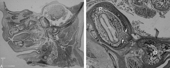
Fig 3.
Photomicrograph of a parasagittal section of the head from an 8-month-old fetus. A, General view. Compared with other structures of the skull base, the otic capsule (frame) appears surprisingly large as it has already attained its adult size. Bony landmarks are F, frontal; Mx, maxillary; Md, mandible; S, sphenoid; C, clavicle; P, petrous; O, occipital; A, atlas. B, Close-up view of the otic capsule. The different layers (o indicates outer; m, middle; i, inner) of the otic capsule are well demonstrated around the basal turn of the cochlea (cbt). A focal dehiscence of the outer layer with protrusion of the middle layer is clearly seen (arrows). The protrusion of the middle layer is directed toward the petro-occipital fissure (pof) and imperceptibly merges with a cartilaginous cap (c) that is distinct from the fibrous tissue occupying the petro-occipital fissure.

