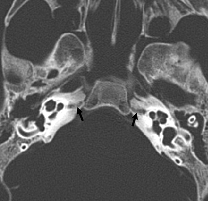
Fig 6.
Transverse MDCT image from a 2-month-old infant specimen. The middle otic layer persists, though tinier than in previous stages. The hypoattenuated focus (arrow) is well demonstrated, spanning the petrous apex, which is now developed and reaching the petro-occipital fissure through a focal cortical interruption.

