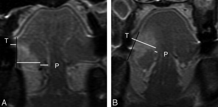
Fig 2.
A, MR image of a 54-year-old man with T2N0 disease shows a vertical black line connecting 2 tumor-mucosa junctions as a reference line. Horizontal white lines are drawn perpendicular to the reference line. Tumor thickness (T) is the sum of both of these horizontal lines and is determined as 10.1 mm (sublingual distance = 0 mm, paralingual distance [P] = 3.8 mm). The elective dissected neck specimen revealed no pathologically positive lymph node. B, MR image of a 41-year-old woman with T3N0 disease demonstrates T of 15.5 mm, sublingual distance of 0 mm, and P of 0.8 mm. Elective dissected neck specimen revealed 1 metastatic node in level III.

