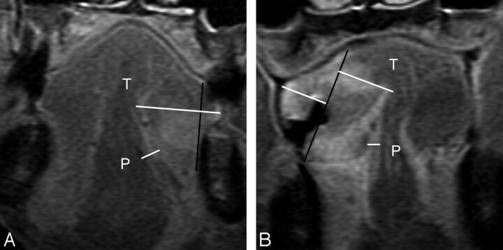
Fig 4.
A, MR image of a 74-year-old man with T4N2b disease, which invaded the mandible, demonstrates a tumor thickness (T) of 19.0 mm, sublingual distance of 0 mm, and paralingual distance (P) of −5.8 mm. Therapeutic neck dissection revealed 9 metastatic nodes in levels I-V. B, MR image of a 61-year-old man with T4N1 disease demonstrates tumor thickness (T) of 27.2 mm, sublingual distance of 0 mm, and paralingual distance (P) of −3.1 mm. The T is the sum of both of these horizontal white lines perpendicular to the reference line. Therapeutic neck dissection revealed 1 metastatic node in level I.

