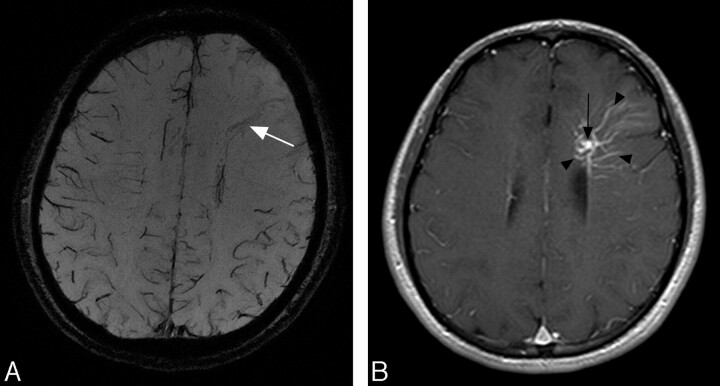
Fig 1.
A 26-year-old man with a DVA. A, MIP image of SWI shows a moderate low-signal-intensity structure corresponding to the draining vein (arrow); however, no low-signal-intensity structures corresponding to medullary veins are seen. Low-signal-intensity structures corresponding to the cortical veins of the other lobes are clearly shown; however, no apparent signals of cortical veins in the left frontal lobe are identified. B, Postcontrast T1-weighted images at 1.5T obtained 2 days after precontrast images reveal the enhanced draining vein (arrow) and medullary veins (arrowheads).

