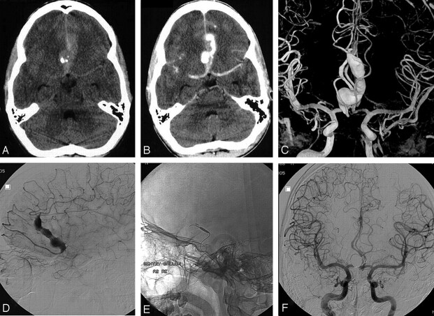
Fig 1.
A 16-year-old boy presented with acute subarachnoid hemorrhage from a giant serpentine anterior cerebral artery aneurysm. A and B, Native (A) and contrast-enhanced (B) CT scans show subarachnoid hemorrhage from a calcified partially thrombosed fusiform aneurysm in the frontal interhemispheric fissure. C, Bilateral internal carotid artery 3D angiography reveals a duplicated right A2 segment with the aneurysm located on the lateral branch. D, Selective angiography of the aneurysm-bearing segment demonstrates branches arising both proximal and distal from the fusiform dilated segment. E, Microballoon for test occlusion. F, Complete exclusion from the circulation after coil occlusion of the proximal part of the lumen. No infarction developed.

