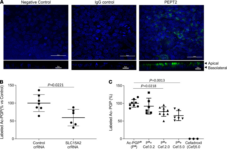
Figure 2. The transport of Ac-PGP in human distal lung epithelial cell line NCl-H441 was inhibited by blocking PEPT2.
The expression of PEPT2 protein (green fluorescence) in polarized NCl-H441 (H441) cells (nuclei, blue fluorescence) was examined by immunohistological staining. Scale bar: 50 μm. Z-stacking images are shown below. Scale bar: 20 μm (A). The transepithelial transport of labeled Ac-PGP (50 ng/ml) in the apical-to-basal directions across PEPT2-knockdown H441 cell monolayers was measured by ESI-LC-MS/MS (B). The transepithelial transport of labeled Ac-PGP (Ac-PGP*, 50 ng/ml) in the apical-to-basal directions across H441 cell monolayers was measured in the presence of PEPT2 inhibitor cefadroxil (0.2 mM, 2.0 mM and 5.0 mM; Cef.0.2, Cef.2.0, and Cef.5.0, respectively) by ESI-LC-MS/MS (C). Data (mean ± SD) are expressed with 3–9 wells per group and were analyzed by Mann-Whitney test or 1-way ANOVA with Tukey’s multiple comparisons post test. crRNA, CRISPR RNA.

