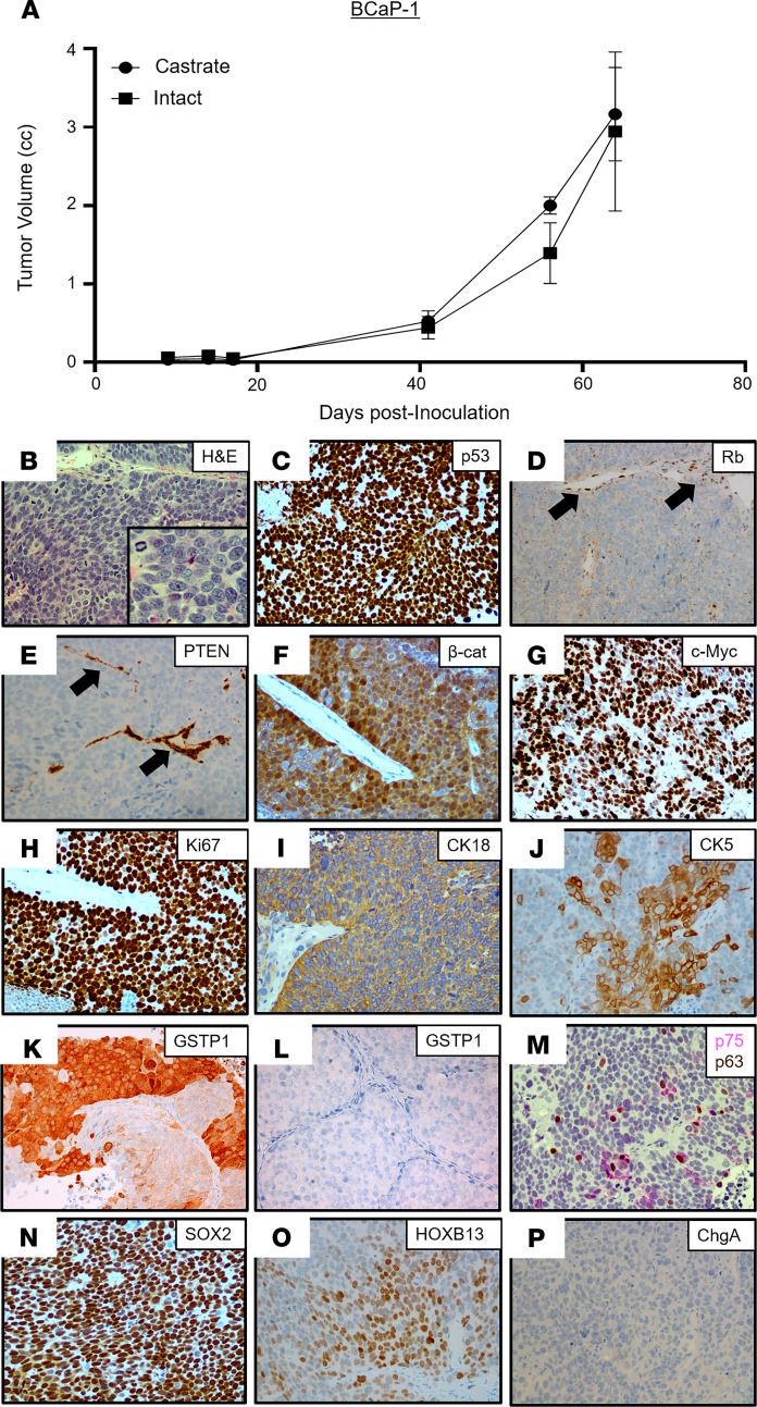
Figure 4. Characteristics of the BCaP-1 PDX.
(A) Growth rate in intact vs. castrated NSG male mice (n = 5). (B) H&E histology (original magnification, ×200; inset [original magnification, ×400]). (C–P) IHC (original magnification, ×200) for (C) p53; (D) Rb (note that endothelial cell nuclei are an internal positive control for staining [black arrows]); (E) PTEN (note that endothelial cells are an internal positive control for staining [black arrows]); (F) β-catenin; (G) c-MYC; (H) Ki67; (I) CK18; (J) focal CK5; (K) GSTP1 in BCaP-1 (positive); (L) GSTP1 in SkCaP-1 (negative control); (M) dual staining for p75 (pink) and p63 (brown); (N) Sox2; (O) HOXB13; and (P) CHGA. PDX, patient-derived xenograft; CHGA, chromogranin A.

