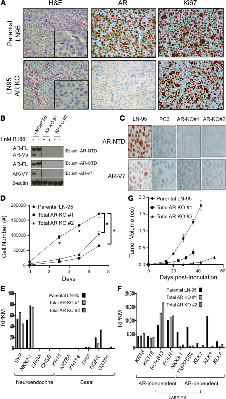
Figure 9. Characterization of LN-95 parental vs.
total AR-KO cells. (A) Left panels are the histology (original magnification, ×200; inset [original magnification, ×400]); middle panels are the AR protein expression (original magnification, ×200); and right panels are the Ki67 expression (original magnification, ×200) of the PDXs. (B) Western blot documentation of the successful KO of AR protein in multiple clones of LN-95 cells. (C) IHC (original magnification, ×200) staining of parental LN-95 cells expressing both full-length AR (AR-FL) and AR variant 7 (AR-V7) vs. AR– PC-3 cells and the AR-KO clones using an N-terminal AR antibody and an AR-V7–specific antibody. (D) In vitro growth of the parental LN-95 cells vs. total AR-KO clones in 10% CS-FBS media, with asterisks denoting a significant difference at the P < 0.05 level. (E) RNA-Seq–based comparison of the expression of NE-specific and basal-specific genes in total AR-KO clones compared with parental LN-95 cells. (F) RNA-Seq–based comparison of the expression of AR-independent and AR-dependent luminal-specific genes in total AR-KO clones compared with parental LN-95 cells (note the significant difference in the magnitude of the y axis between panels). (G) In vivo growth of the total AR-KO clones vs. the parental LN-95 in castrated hosts. LN-95, LNCaP-95.

