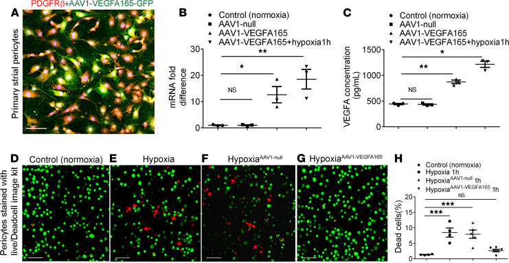
Figure 5. AAV1-HRE-VEGFA165 transfer to pericytes significantly promotes pericyte survival under hypoxic conditions.
(A) AAV1-HRE-VEGFA165-GFP successfully infected a cochlear pericyte cell line with a multiplicity of infection (MOI) of 1 × 10⁵ when imaged 48 hours later. (B) Real time-quantitative PCR shows significant upregulation of Vegfa mRNA (n = 3 for each group, *P < 0.05, and **P < 0.01 by 1-way ANOVA). (C) ELISA shows high production of VEGFA protein in an AAV1-HRE-VEGFA165 gene-infected pericyte relative to an AAV1-GFP null-infected pericyte (n = 3 for each group, *P < 0.05, and **P < 0.01 by 1-way ANOVA). (D–G) Distribution of live (green) and dead pericytes under hypoxia as measured with a Live/Dead Cell Viability Assay Kit (MilliporeSigma). (H) Statistical analysis shows increased pericyte survival (green) under hypoxic conditions when pericytes were pretransfected with AAV1-HRE-VEGFA165 for 48 hours (n = 4 for control, n = 4 for hypoxia 1h, n = 5 for AAV1-GFP hypoxia 1h, n = 6 for AAV1-VEGFA165 hypoxia 1h, and ***P < 0.001 by 1-way ANOVA). Data are presented as mean ± SEM. Scale bars: 100 μm.

