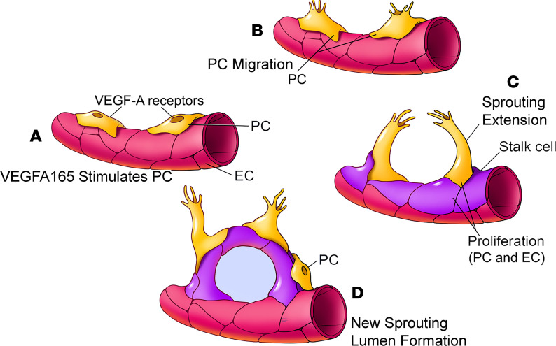
Figure 9. A model of VEGFA165-controlled angiogenesis in the cochlea.
(A and B) VEGFA165 induces pericytes to migrate. (C) The pericytes “sense” the VEGFA165 diffusing from vessels and align along the VEGFA165 gradient to form a “sprout.” The proliferation of ECs behind the tip cells drives the precapillary to elongate. (D) Newly proliferated pericytes on the new branch tube establish focal contacts with ECs.

