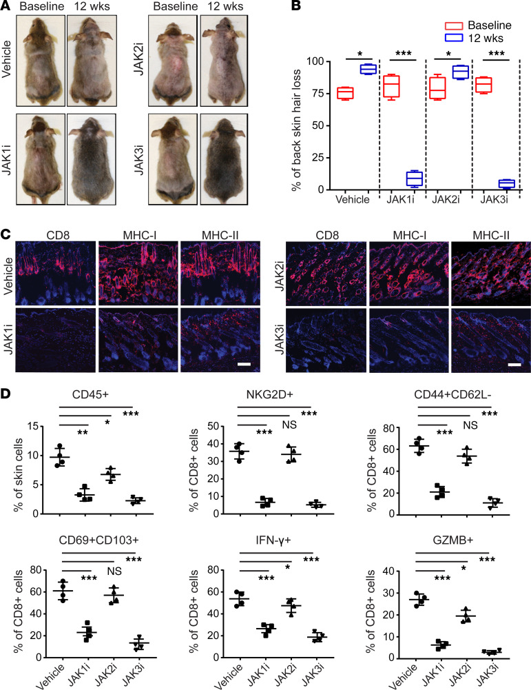
Figure 5. Reversal of AA with topical JAK1- or JAK3-selective inhibitor treatment.
C3H/HeJ mice with long standing AA were treated topically with INCB039110 (JAK1i), CEP-33779 (JAK2i), or PF-06651600 (JAK3i) or control daily for 12 weeks, in cohorts of 4 mice per group. (A) Representative images of individual JAK inhibitor– or vehicle-treated C3H/HeJ mice before or after 12 weeks’ treatment. (B) Percentage of dorsal hair loss or regrowth is shown before and after treatment. The box plots depict the minimum and maximum values (whiskers), the upper and lower quartiles, and the median. The length of the box represents the interquartile range. *P < 0.05, ***P < 0.001 (unpaired Student’s t test). (C) Representative immunofluorescence images of skin sections from JAKi- or vehicle-treated mice, stained with anti-CD8, anti–MHC class I, or anti–MHC class II mAbs. Scale bar: 100 μm. (D) Percentages of skin infiltrating CD45+ leukocytes, NKG2D+CD8+ T cells, CD44+CD62L–CD8+ T cells, CD103+CD69+CD8+ T cells, IFN-γ–producing CD8+ T cells, as well as GZMB- or PRF1-producing CD8+ T cells within indicated populations within the skin after JAK inhibitor treatment. *P < 0.05, **P < 0.01, ***P < 0.001 (1-way ANOVA). Two replicate experiments were performed for a total of 8 mice per group.

