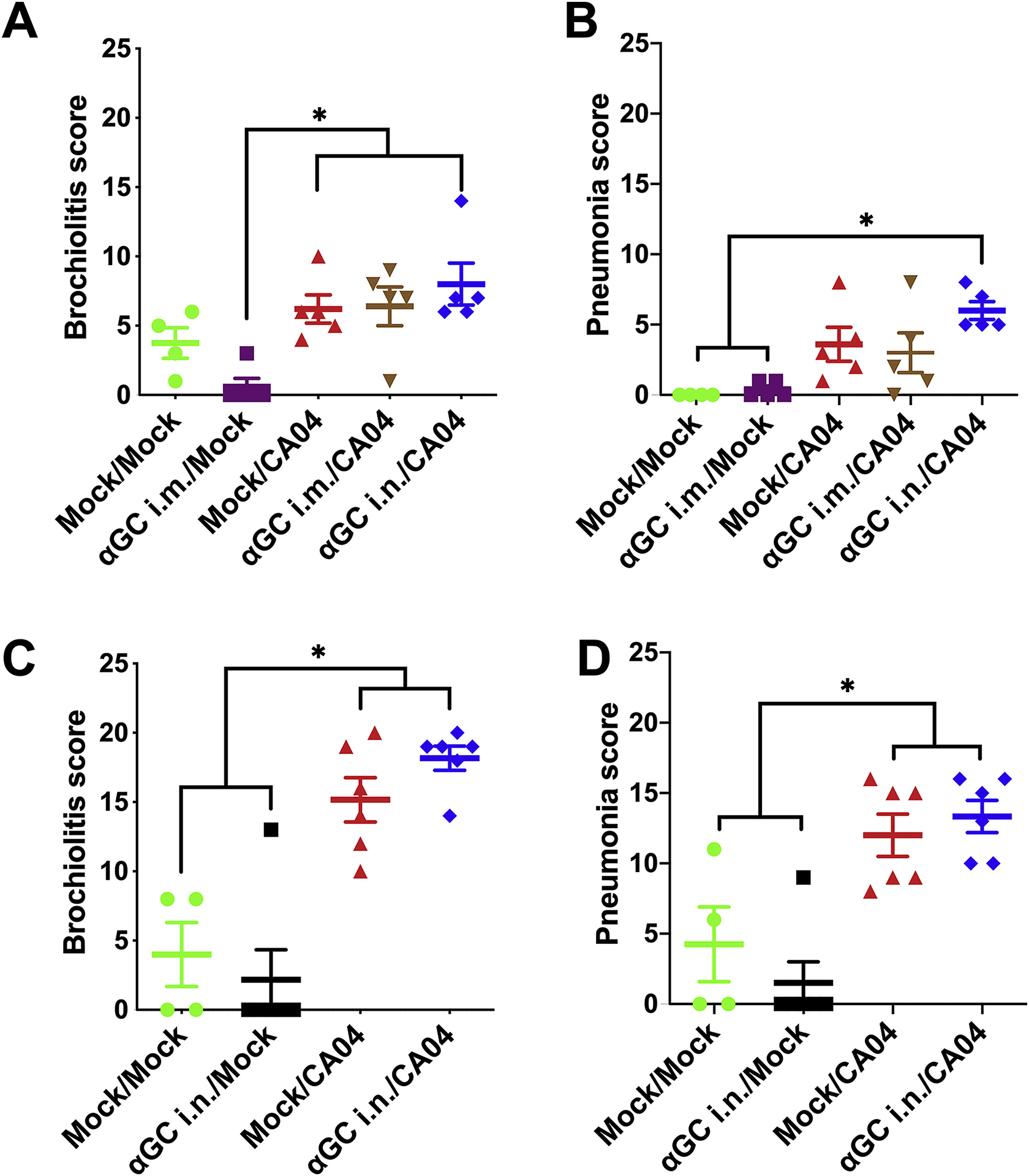
Figure 3.

Therapeutically activating iNKT cells prior to IAV infection does not alter lung pathology. Pathology was assessed by evaluating H&E stained lung sections from pigs administered α-GalCer (αGC) i.m. or i.n. 9 days prior to infection (A & B) or i.n. 2 days prior to infection (C & D). (A & C) Bronchiolitis score; (B & D) Pneumonia score. Data are presented as mean ± SEM. Data were analyzed using the SAS PROC GLIMMIX procedure and differences between groups were analyzed using Tukey’s test, *P < 0.05.
